Neurologic examination (lesion localization)
IVDD (Intervertebral disk disease)
- Cervical – Ventral slot
- Thoraco-lumbar – Hemilaminectomy
- Lumbosacral – Dorsal laminectomy
Neoplasia (cancer)
Wobblers (Cervical spondylomyelopathy)
Atlanto-axial instability (AA luxation)
Spinal fracture fixation
Nerve/muscle biopsy
Spinal tap (CSF tap)
Myelogram


Soft Tissue Surgery
Exploratory celiotomy (abdominal surgery)
- Cholecystectomy (gall bladder removal)
- Portosystemic shunt attenuation (PSS)
- Gastropexy (prophylactic and fixation for GDV)
- Gastrodilatation Volvulus (GDV) -gastropexy
- Common bile duct stenting
- Intestinal resection with anastomosis
- Subtotal colectomy
- Colopexy
- Thoracic duct ligation/cisterna chyli ablation/pericardectomy (for chylorthorax)
Hernia repair
- Perineal herniorrhaphy
- Gastroesophageal herniorrhaphy (hiatal hernia repair)
- Diaphragmatic herniorrhaphy
- Inguinal herniorrhaphy
- Scrotal herniorrhaphy
Ear disease
- Ventral bulla osteotomy (VBO)
- Total ear canal ablation with lateral bulla osteotomy (TECA-LBO)
Eye/eyelid disease
- Enucleantion/Exenteration
- Cherry eye repair (third eyelid gland)
- Entropion
Head and Neck
- Brachycephalic syndrome surgery
- Sialocelectomy (removal of salivary glands)
- Arytenoid lateralization (laryngeal tieback for Laryngeal Paralysis)
Perineal/anal surgery
- Rectal polyps removal
- Anal sacculectomy
- Caudectomy (for screw tails primarily English bulldogs)
Chest/Heart disease
- Patent duct arteriosus ligation (PDA)
- Persistent right aortic arch (PRAA)
- Lung lobe torsion
- Bleb/bulla – pneumothorax
Skin procedures
- Mesh grafts
- Skin flaps
- Advancement flaps
Urinary/reproductive disease
- Ectopic ureters
- Cystotomy
- Urethrostomy (perineal, prescrotal, and scrotal)
- Urethral prolapse
- Caesarean section
- Cryptorchidectomy
- Pyometra
Oncologic Surgery
Thoracotomy
- Lung tumors (lung lobectomies)
- Pericardectomies
Mandibulectomies
Maxillectomies
Large tumor resections (skin) with/without skin flap or mesh graft
Amputations
- Forelimb amputation
- Rear limb amputation
- Digit amputation
- Tail amputation
TECA-LBO (total ear canal ablation with lateral bulla osteotomy)
Exploratory celiotomy
- Intestinal resection anastamosis
- Nephrectomy
- Adrenalectomy
- Pancreatectomy
- Colectomy
- Splenectomy
- Hilar resection/liver lobectomy
- Bladder resection
Thyroidectomy/parathyroidectomy
Mastectomy/mammectomy
Chest wall reconstruction
Venous access port placement (VAP)
Orthopedic Surgery
Lameness evaluation
Fracture repair – all bones
- Appendicular
- Axial
Cranial cruciate rupture
- TPLO (Tibial Plateau Leveling Osteotomy)
- Extracapsular repair (Extra cap)
- Meniscectomy
- Arthrotomy
Elbow Dysplasia
- Ununited anconeal process (UAP)
- Fragmented medial coronoid process (FCP)
- Elbow incongruency
- Osteochondrosis (OCD)
Arthrodesis (joint fusion)
- Carpus
- Tarsus
- Elbow
- Shoulder
Joint Disease
- Angular limb deformities – corrective osteotomies
- Subluxation/Luxation – Reduction
- Arthrocentesis (joint tap)
- Joint injections
- Joint lavage
Ligament/tendon disease
- Achilles tendon rupture
- Biceps tendon
- Supraspinatus
- Infraspinatus
Medial/lateral patella luxation
- Tibial tuberosity transposition
- Block recession
- Retinacular imbrication/release
- Corrective osteotomies – if severe angular deformity
Hip Disease
- Femoral head and neck ostectomy
- Triple Pelvic Osteotomy (TPO)
- Juvenile pubic symphysiodesis (JPS)
- Joint injections
Shoulder Disease
- Osteochondrosis dessicans (OCD)
- Medial shoulder instability
- Shoulder luxation
- Sacroiliac Luxation


Cranial Cruciate Surgery
Causes & Symptoms
Stability of the stifle (knee) is provided by several different structures:
- Cruciate ligaments
- Cranial cruciate ligament (CrCL)
- Caudal cruciate ligament (CdCL)
- Medial and lateral collateral ligaments
- Joint capsule
- Menisci
- Peri-articular muscles
Instability can cause pain, lameness, and osteoarthritis. The number one reason for lameness in the knee is a ruptured CrCL.
The origin of the CrCL is the medial aspect of the lateral condyle, and it inserts on the cranial aspect of the tibial plateau. It prevents cranial translation of the tibia (“drawer motion”), excessive internal rotation and hyperextension of this joint. There are 2 parts of the CrCL which makes it complex: “craniomedial band” (taught flexion and extension) and “caudolateral band” (lax in flexion).
CrCL rupture is the most common reason for rear limb lameness in the dog. It is more common in large and giant breed dogs but is also commonly recognized in smaller breeds of dogs and cats. The three most common implemented causes for rupture of the CrCL include:
Supra-physiologic loads (athletic dogs in which hyperextension or severe internal rotation occurs)
Ligament degeneration (occurs in older dogs and sedentary life style)
Other causes (conformational abnormalities, joint inflammation, primary degenerative changes, and immune mediated causes).
Clinical signs can be very subtle from barely lame (common with partial tear or hyperextension) to non-weight bearing lameness (full tear with meniscal tear). Most patients with CrCL rupture present to your regular veterinarian for acute non-weight bearing lameness that may improve initially, then worsen again. Some ruptures may go unnoticed or undetected and can present as a chronic, insidious, or intermittent lameness. However, commonly with a CrCL rupture there is stifle (knee) pain and joint effusion palpated on orthopedic exam.
Diagnosis
An orthopedic examination is needed to find or confirm the CrCL rupture. Joint effusion (swollen joint) and medial buttress (fibrous tissue on the inner aspect of the stifle) are almost always present on orthopedic exam when a CrCL rupture is present. Most dogs have cranial drawer (see video below), but with chronic ruptures and severe ruptures, the stifle may already be sitting in the cranial drawer position, therefore, this orthopedic exam finding may be absent.
Most dogs have cranial drawer (see video below), but with chronic ruptures and severe ruptures, the stifle may already be sitting in the cranial drawer position, therefore, this orthopedic exam finding may be absent.
Cranial translation of the tibia (tibial thrust) is always found on the orthopedic exam when a CrCL rupture is present. As you can see in the following video, this is an abnormal forward movement of the tibia that does not occur unless a rupture/tear has occurred.
Other findings that could be found on orthopedic exam include but are not limited to:
- Meniscal click
- Crepitus
- Pain on hyperextension
- Internal rotation
Radiographs are also used to help diagnose a CrCL rupture. Radiographs are used along with the orthopedic exam to confirm the diagnosis of CrCL rupture. Common radiographic changes associated with CrCL rupture include:
- Joint effusion
- Evidence of osteoarthritis (on the patella, trochlear ridges and tibial plateau)
- The stifle (knee) in the cranial drawer position
Radiographs with specific positions and angles will probably need to be performed by your surgeon to help plan for the surgery performed.
Treatment
Treatment for this disease process is easily broken down into two categories: conservative or surgical management. Conservative management consists of pain medications, anti-inflammatories and strict cage confinement, allowing the body ample time to heal and stabilize. There are several different surgical techniques to stabilize the stifle, meaning there is not one specific and ideal way to correct the instability. We offer 2 surgical techniques to stabilize your pet’s stifle.
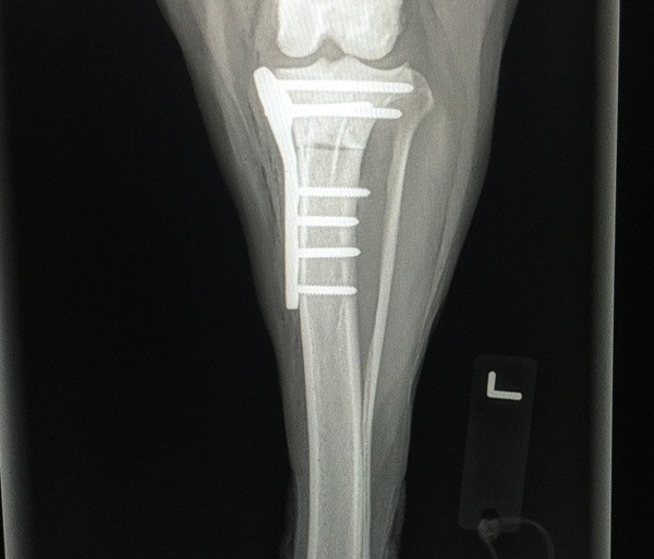
The first is the Tibial Plateau Leveling Osteotomy (TPLO). This is more invasive than the second option, but has been proven to be far superior with better clinical results than other procedures. The TPLO involves making a circular cut (osteotomy) in the tibia and rotating the proximal aspect of the cut to flatten the plateau of the tibia. The bone plate is then placed to hold the top and bottom parts of the cut in place.
The second procedure is the extracapsular technique. This procedure involves placing a non-absorbable suture in the same orientation as the ruptured CrCL which serves as a prosthetic ligament, allowing the body and the suture to stabilize the knee. It can be performed on any size dog or cat, but it is normally a better option to perform in smaller dogs due to the fact of the higher chance of failure in larger dogs. Despite the surgical treatment, an arthrotomy (exploration of the joint) will be performed to visualize the tear, investigate the integrity of the meniscus, lavage the joint, debride the remnants of the CrCL, and to visualize other structures in the diseased joint.
Post-Surgical Care
Strict cage confinement is imperative for any treatment performed, but it is very important for the surgical treatments. It normally takes 10-12 weeks for normal healing to occur depending on the surgical technique used. Several recheck visits with radiographs are necessary for determination of healing and to evaluate the surgical site. For more information on diagnosing, consultation, and treatment of ruptured CrCL, please email or call our office.


![]()
Board-certification by the American College of Veterinary Surgeons
What does it mean?
- Four years of advanced training in surgery beyond the Doctor of Veterinary Medicine Degree
- Experience in the development of new surgical treatments
- Rigorous examination by the American College of Veterinary Surgeons to ensure competency in advanced surgical techniques
- Certifies that a veterinarian is a surgical specialist