Causes & Symptoms

Stability of the stifle (knee) is provided by several different structures:

  • Cruciate ligaments
    • Cranial cruciate ligament (CrCL)
    • Caudal cruciate ligament (CdCL)
  • Medial and lateral collateral ligaments
  • Joint capsule
  • Menisci
  • Peri-articular muscles

Instability can cause pain, lameness, and osteoarthritis. The number one reason for lameness in the knee is a ruptured CrCL.
The origin of the CrCL is the medial aspect of the lateral condyle, and it inserts on the cranial aspect of the tibial plateau. It prevents cranial translation of the tibia (“drawer motion”), excessive internal rotation and hyperextension of this joint. There are 2 parts of the CrCL which makes it complex: “craniomedial band” (taught flexion and extension) and “caudolateral band” (lax in flexion).
CrCL rupture is the most common reason for rear limb lameness in the dog. It is more common in large and giant breed dogs but is also commonly recognized in smaller breeds of dogs and cats. The three most common implemented causes for rupture of the CrCL include:

Supra-physiologic loads (athletic dogs in which hyperextension or severe internal rotation occurs)
Ligament degeneration (occurs in older dogs and sedentary life style)
Other causes (conformational abnormalities, joint inflammation, primary degenerative changes, and immune mediated causes).

Clinical signs can be very subtle from barely lame (common with partial tear or hyperextension) to non-weight bearing lameness (full tear with meniscal tear). Most patients with CrCL rupture present to your regular veterinarian for acute non-weight bearing lameness that may improve initially, then worsen again. Some ruptures may go unnoticed or undetected and can present as a chronic, insidious, or intermittent lameness. However, commonly with a CrCL rupture there is stifle (knee) pain and joint effusion palpated on orthopedic exam.

Diagnosis

An orthopedic examination is needed to find or confirm the CrCL rupture. Joint effusion (swollen joint) and medial buttress (fibrous tissue on the inner aspect of the stifle) are almost always present on orthopedic exam when a CrCL rupture is present. Most dogs have cranial drawer (see video below), but with chronic ruptures and severe ruptures, the stifle may already be sitting in the cranial drawer position, therefore, this orthopedic exam finding may be absent.

Most dogs have cranial drawer (see video below), but with chronic ruptures and severe ruptures, the stifle may already be sitting in the cranial drawer position, therefore, this orthopedic exam finding may be absent.

Cranial translation of the tibia (tibial thrust) is always found on the orthopedic exam when a CrCL rupture is present. As you can see in the following video, this is an abnormal forward movement of the tibia that does not occur unless a rupture/tear has occurred.

Other findings that could be found on orthopedic exam include but are not limited to:

  • Meniscal click
  • Crepitus
  • Pain on hyperextension
  • Internal rotation

Radiographs are also used to help diagnose a CrCL rupture. Radiographs are used along with the orthopedic exam to confirm the diagnosis of CrCL rupture. Common radiographic changes associated with CrCL rupture include:

  • Joint effusion
  • Evidence of osteoarthritis (on the patella, trochlear ridges and tibial plateau)
  • The stifle (knee) in the cranial drawer position

Radiographs with specific positions and angles will probably need to be performed by your surgeon to help plan for the surgery performed.

Treatment

Treatment for this disease process is easily broken down into two categories: conservative or surgical management. Conservative management consists of pain medications, anti-inflammatories and strict cage confinement, allowing the body ample time to heal and stabilize. There are several different surgical techniques to stabilize the stifle, meaning there is not one specific and ideal way to correct the instability. We offer 2 surgical techniques to stabilize your pet’s stifle.

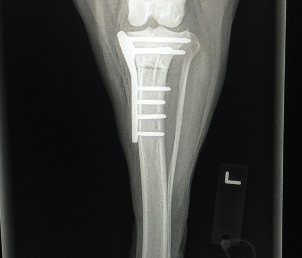
The first is the Tibial Plateau Leveling Osteotomy (TPLO). This is more invasive than the second option, but has been proven to be far superior with better clinical results than other procedures. The TPLO involves making a circular cut (osteotomy) in the tibia and rotating the proximal aspect of the cut to flatten the plateau of the tibia. The bone plate is then placed to hold the top and bottom parts of the cut in place.

The second procedure is the extracapsular technique. This procedure involves placing a non-absorbable suture in the same orientation as the ruptured CrCL which serves as a prosthetic ligament, allowing the body and the suture to stabilize the knee. It can be performed on any size dog or cat, but it is normally a better option to perform in smaller dogs due to the fact of the higher chance of failure in larger dogs. Despite the surgical treatment, an arthrotomy (exploration of the joint) will be performed to visualize the tear, investigate the integrity of the meniscus, lavage the joint, debride the remnants of the CrCL, and to visualize other structures in the diseased joint.

Post-Surgical Care

Strict cage confinement is imperative for any treatment performed, but it is very important for the surgical treatments. It normally takes 10-12 weeks for normal healing to occur depending on the surgical technique used. Several recheck visits with radiographs are necessary for determination of healing and to evaluate the surgical site. For more information on diagnosing, consultation, and treatment of ruptured CrCL, please email or call our office.

max-ward-tplo